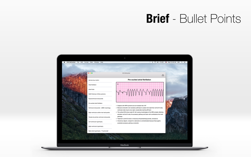
Simple and Basic. To master ECG interpretation takes time and effort, however this app is designed to be concise and focused on only what you need to know, yet very thorough.
Knowing the basic parts of an ECG tracing will lay a good foundation for everything else that is to come. The different waves, complexes and intervals need to be ingrained in your brain. How many seconds is a full ECG tracing and how much time does each big and each little box represent?
This app is not for learning the crazy things such as the different P wave morphologies that occur with atrial enlargements and ectopic atrial rhythms... just know what the normal P wave looks like and what it represents. Similar concept for the other parts of the ECG.
You learn things like this by using the ECG Refresher app:
Wave: A positive or negative deflection from baseline indicates a specific electrical event. The waves on an ECG include the P wave, Q wave, R wave, S wave, T wave and U wave.
Interval: The time between two specific ECG events. The intervals that are commonly measured on an ECG include the PR interval, QRS interval (also called QRS duration), the QT interval and the RR interval.
Segment: The length between two specific points on the ECG which are supposed to be at the baseline amplitude (not negative or positive). The segments on an ECG include the PR segment, ST segment and the TP segment.
Complex: The combination of multiple waves grouped together. The only main complex on the ECG is the QRS complex.
Point: There is only one point on the ECG termed the J point which is where the QRS complex ends the ST segment begins.
The main part of the ECG contains a P wave, QRS complex, and T wave which will each be explained individually in this tutorial as will each segment and interval.
The P wave indicates atrial depolarization. The QRS complex consists of a Q wave, R wave and S wave. The QRS complex represents ventricular depolarization. The T wave comes after the QRS complex and indicates ventricular repolarization. Below is a normal QRS complex with the individual parts labeled and a normal full 12-lead ECG:


